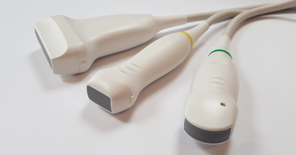
Did you know that certain ultrasound probes (transducers) are better at certain applications than others? It’s important to make sure you select the correct probe for the job in hand. Probes are expensive and many veterinary sites get by with one ‘jack of all trade’ microconvex probe for both abdominal and cardiac images, but ultimately this is a compromise, and I would recommend having additional probes if both budget and scanner allow. Here’s a very brief rundown on the recommended probe types for small animal veterinary applications in the modern practice:
Microconvex Probe
Small, curved footprint probe, ideal for general abdominal scans in companion animals, typically these probes are mid to high frequency and offer great image quality in the near to mid field, however the deeper the image the poorer the images as the beams diverge and the scanner has to ‘fill in’ the gaps between the beams. If the structure you are looking at is quite deep, try to scan from a different location and make the structure of interest more superficial, allowing it to be the in the ‘sweet spot’ of the probe and allowing use of the higher frequency range of the probe, theoretically allowing better image quality/resolution. If the abdomen is quite large and the microconvex probe struggles, I would recommend the…
Convex Probe
Large footprint curved probe, typically operating in the mid to low frequency ranges meaning it is optimised of deeper abdominal studies. It’s not as wide angle as the microconvex probe which means that the beams diverge less the deeper the study and the image quality in these studies is significantly better than the microconvex probe even though it is a lower frequency probe. It’s often overlooked due to its bigger physical size but is recommended if you see a lot of large breeds or obese cases.
Linear Probe
A short straight probe, usually about 4cm in length and great for most superficial soft tissue studies (up to 6 or 7 cm). The linear probes are usually higher frequency probes, which offer higher image resolution (but less penetration) making them a superb probe for imaging GI tract, spleens, kidneys, Adrenals, lymph nodes, vascular and arterial studies, MSK work, even scanning eyes. A linear probe really is worth getting for improving imaging on any superficial soft tissue.
Phased Array Probe
Small square footprint probe providing an image from a ‘point source’ designed for cardiac scanning where they enable superb views of the heart in both long and short axis with minimised rib shadowing. The phased array probes are available in various frequency ranges to suit differing sizes of patients, they also offer faster frames rates and operate the cardiac imaging modes of the scanners that don’t operate with the abdominal probes, such as Continuous Wave Doppler (CW) and Tissue Doppler/Velocity imaging (TDI/TVI) as well as the more advanced cardiac functions. A lot of ultrasound users in vet practices are limited in their cardiology imaging by only having the microconvex probe, so if you are looking to advance your cardiology skills it is worth making sure the scanner you select has the ability to use the phased array type of probe.
We will shortly be adding a ‘probes’ page to the website, but if you have any questions about selecting the correct probe for a specific scan or would like help with a recommendation for purchasing additional probes or to book a training session on your ultrasound scanner then please contact us at info@ultrasoundscannertraining.co.uk or fill out the contact form.
