The frame rate that you use when scanning will have an effect on the image attained. Depending on how wriggly the patient is or if they are panting, too slow a frame rate will potentially result in a blurred looking image, too high a frame rate and you could be losing out on image quality. Frame rate is an important consideration in all scans and should be tailored to suit the scan being carried out, i.e. abdominal or cardiac.
There are a few ways of adjusting the frame rate:
- The simplest is to adjust the depth you are scanning at, if you are looking deep into the body the frame rate will be quite low(measured in Hz on the scanner), reducing the depth being scanned (as long as you can still see the structure of interest) should increase the frame rate. To allow you to image a structure at a reduced depth you may need to move the probe so that the structure you are scanning lies closer to the probe, this has other advantages too, you will be able to use a higher frequency potentially providing a better image.
- Decrease the size of the sector width, or, how wide the image is, most scanners will have a control on to allow this. If the frame rate is too low, make the sector narrower. Sme scanners will also allow you to change the angle of the narrowed sector too.
- A third option is a little more involved and depending on the make of system you use this is the persistence or correlation, although adjusting this will not change the frame rate, it will give the illusion of doing so. Images with high persistence can look ‘blurred’. If this is the case, reduce the persistence and the image should seem to ‘speed up’. This is useful to keep up with the animals breathing, panting or other movements. Be careful though as reducing it too low can make the image look quite coarse.
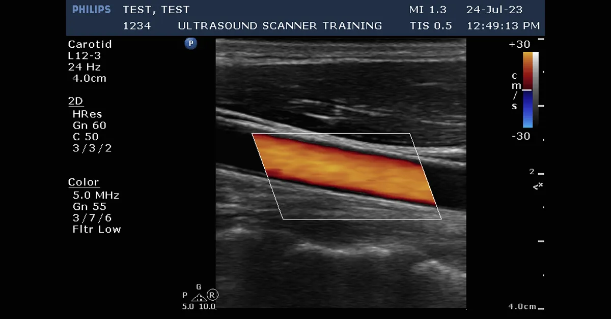
At the end of the day adjusting the frame rate is a bit of a balancing act. Try a combination of adjustments and see what happens to the image. If you have a really fast frame rate, the chances are you’ll not be able to see all the frames at full speed, use cine loop and slow the review speed down, you’ll start to see all of the frames and more detail too. This is a great function in cardiology. When using colour doppler try to keep the size of the colour box or ‘RoI” sensible, if its big the frames rates will be lower, in the example image you can see that I’ve kept the colour box only around the area to be sampled and the frame rate is quite good at 24Hz (shown in the scan information at top left of image).
If you need help understanding your ultrasound scanner or have new staff who require system training, please call me on 07795322354 or email info@ultrasoundscannertraining.co.uk. Head to our other blog pages for further information, hints and tips.