Home > Services
Are you looking to get the most out of your ultrasound scanner?
We provide ultrasound scanner training at various levels and aim to improve your understanding of the principles of ultrasound. We will develop your skills driving your equipment, enabling you to optimise images so you can provide a fast and accurate diagnosis. It’s important to note that our training helps you ‘drive’ your scanner and is complementary to the various diagnostic training courses available.
Foundation Training
General Training
Advanced Training
Ultrasound scanner training for every level.
We understand that every practice is different. New staff need an introduction to your particular ultrasound equipment, experienced staff sometimes need top-up training. We’ve got you fully covered with whatever level of training you need. Just ask – we’ll get your whole team working efficiently together and confident in their decision making.
Ever wondered what that button does?
It’s well known that many users of ultrasound equipment barely scratch the surface of their scanners true potential, only using a few controls, in some cases adjusting controls just to ‘see what happens’ without a real understanding of what to adjust and, equally as importantly, when. Scanners from different manufacturers often have alternative terminology for the same thing, we will go through these and bring clarity to the controls. Now is the time to unlock the potential and see what your scanner can really do!
- Creating a straightforward routine to follow from system power up to scanning for each patient.
- Cover the correct patient preparation to enable the best signal for image quality.
- Choosing the correct probe for each application, different probe types have applications that they’re best suited to.
- Making sure the presets on your scanner are suitable and selecting the correct one for the scan to be performed, refining and creating your own presets.
- Understanding and adjusting image orientation and how it’s displayed on screen.
- Reviewing and using the various software filters that your scanner may have.
- Taking routine measurements and utilising the analysis and reporting capability of your scanner.
- Use of annotation options on your scanner – text lists for each preset type and free text.
- Image storage and archiving.
- Starting to scan and getting to grips with the main controls to give you the best image resolution quickly and efficiently:
- Depth and zoom - how to best display the structure being investigated.
- Focus – how to alter the focus position, the benefits and drawbacks of adding extra focal zones or increasing the size of focal area.
- Frequency – understanding the basic principals of imaging frequency, when and how to adjust.
- Gain – getting to grips with both the individual gain controls (Time Gain Compensation) and the Overall Gain controls plus the lateral gain controls if your system has this ability.
- Refining and optimising the image further with the more advanced controls including:
- Dynamic range.
- Gray maps.
- Sector width or scan angle for curved and phased probes.
- Image width and extended field of view (trapezoidal or virtual convex) for linear probes.
- Persistence (also referred to as frame averaging or correlation depending on scanner brand).
- The correct use of Tissue Harmonic Imaging (THI/CHI/PHI).
- Line density and the effects on frame rates.
Training sessions can be tailored to suit the needs of your team on your own equipment, going through the controls of the following imaging modes routinely used in everyday applications:

B Mode Imaging
Sometimes referred to as 2D imaging. This is the standard black and white view that all scanners, from the most basic will provide.
Do you need help understanding ultrasound imaging modes? When you book training with us we will take you through all the settings of your ultrasound equipment in a friendly, easy to understand way.

M Mode Imaging
For viewing and analysing the function of the heart against time. This is the mode used for a wide range of cardiac measurements. Some systems with a cardiac bias also have a feature known as Anantomical M Mode which allows more freedom of the cursor used to choose the section of the heart to be viewed against time.

Colour Flow Doppler
Applies colour to blood flow through the organs of the body allowing the operator to visualise the bloodflow and pinpoint issues. Colour Doppler is used to qualify blood flow but not quantify it.
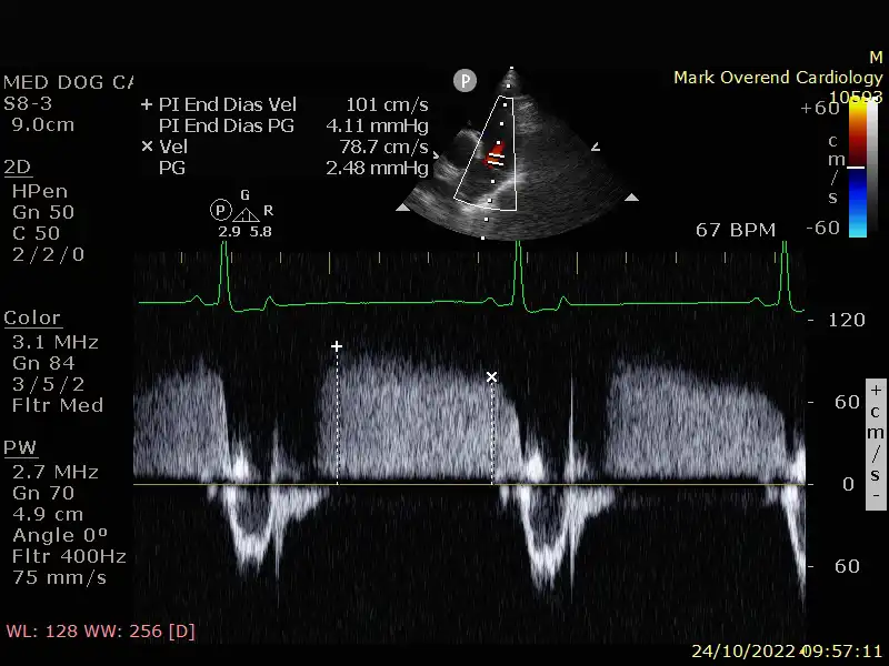
Pulsed Wave Doppler
Allows the operator to measure specific blood velocities, used both in abdomen and cardiac applications.

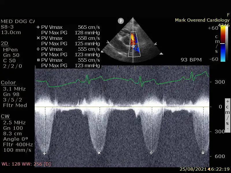
Continuous Wave Doppler
Similar to Pulsed Wave but used for faster blood velocities typically found in issues with the heart such as Mitral Valve Regurgitation or other 'jets'. This is a very useful tool in diagnosing cardiac insufficiencies.
Modular training sessions
Refresher courses
Learn new techniques
We can help with your ultrasound.
- Become more assured using your ultrasound scanner and all its features.
- Gain confidence knowing which probe to select for your applications.
- Improve your skills and techniques for fast and reliable diagnosis.